This article details grossing for neoplastic gastrectomy or esophagectomy specimens.
Initial orientation
Before beginning grossing, take time to orient these specimens properly. Key anatomical landmarks include:
Gastroesophageal junction (GEJ)
- The GEJ is the point where the tubular esophagus meets the stomach, regardless of the type of epithelium at the junction
Esophagus
- Lacks serosa – look for a roughened, dull outer surface (adventitia)
Greater omentum (total gastrectomy only)
- Arises from the greater curvature of the stomach, draping downward and connecting to the transverse colon
Lester omentum
- Arises from the lesser curvature and connects to the liver
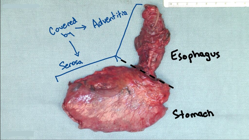
Distal esophagectomy with proximal stomach
- Esophagus is covered in adventitia and does not have a serosal covering (it will have a roughened texture/appearance compared to the stomach)
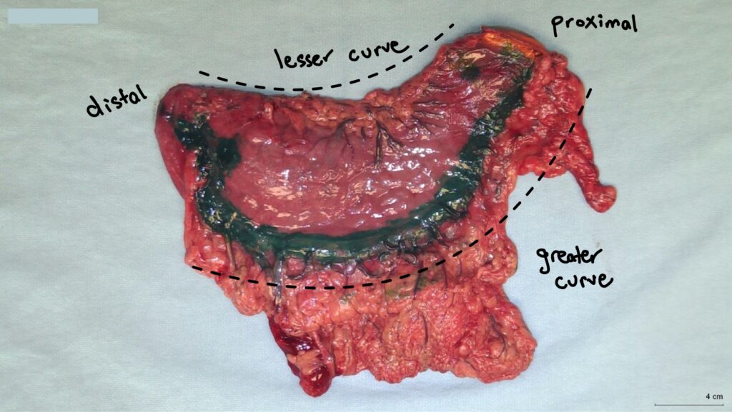
Total gastrectomy with green ink along greater curve
- Greater omentum extends off the greater curve of the stomach
- Lesser omentum extends off the lesser curve of the stomach
- Omental margins on a gastrectomy are the only radial margins
Inking Margins
The distal margin may have already been inked prior to opening. If not already done, ink the adventitial and radial margins before proceeding:
- Esophageal (inked blue in following photo)
- Total gastrectomy: ink along the lesser and greater curves (red lines in following photo) to designate radial omental margins
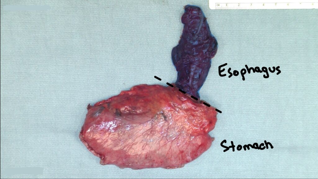
Esophageal adventitial margin inked blue
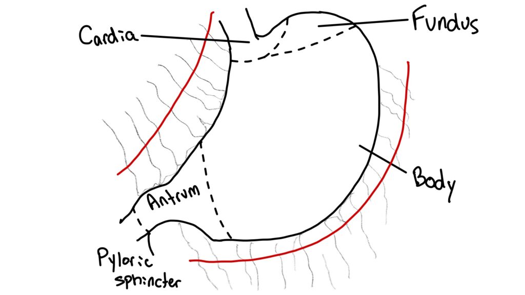
Ink along the red lines (where omentum has been cut) to indicate radial omental margins.
Esophageal vs Gastric Tumor?
One of the the main goals of the gross description is to classify the tumor as esophageal or gastric, as this determines which staging system to use. If the tumor involves only the esophagus or only the stomach without crossing or involving the gastroesophageal junction, this is straightforward.
However, if both structures are involved, evaluating the tumor mid-point will determine the origin of the tumor.
Cancer involving the GEJ with the tumor midpoint greater than 2 cm into the proximal stomach is classified as a gastric tumor.
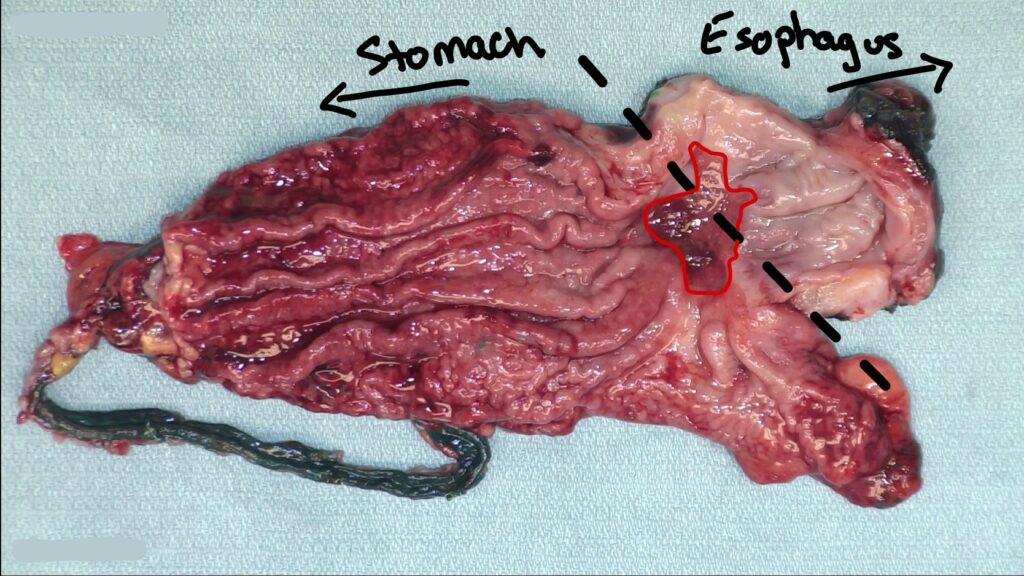
In this specimen, the tumor involves both the esophageal and gastric mucosa and the midpoint is less than 2 cm into the gastric mucosa → esophageal tumor.
One last note on determining esophageal vs. gastric tumors. A tumor of the cardia or proximal stomach without involvement of the GEJ, even if the tumor midpoint is less than 2 cm into the proximal stomach, is still classified as a gastric tumor.
Landmarks to report:
For total gastrectomy specimens, Describe the tumor location using
- Curvature: Greater vs lesser curve
- Region: Cardia, fundus, body, antrum
- Wall: Anterior vs posterior
https://documents.cap.org/documents/Stomach_4.4.0.0.REL_CAPCP.pdf?_gl=1*9cq86q*_ga*MTc2OTc4NjM4MS4xNzQ3MzEzNjQw*_ga_97ZFJSQQ0X*czE3NTMyODQ0NjQkbzQkZzAkdDE3NTMyODQ0NjkkajU1JGwwJGgw
Structuring the Gross Description
- What was received
- Total gastrectomy: Includes stomach, distal esophagus, lesser and greater omentum
- Distal esophagectomy/partial gastrectomy: Includes distal esophagus, proximal stomach, and lesser omentum
Include:
- Lengths along lesser and greater curves
- Open circumferences at proximal and distal margins
- Describe the primary pathology
- Provide dimensions
- Describe the appearance (eg ulcerated, heaped up borders with ulcerative center etc)
- Localize within the stomach/esophagus
- Assess depth of invasion
- Measure distance to margins
→ Relevant margins to include for these specimens are:
- Proximal
- Distal
- Radial (lesser and greater omental margins are the radial margins for a total gastrectomy; lesser omental and the esophageal adventitial margin are radial margins for a esophagectomy)
- Stapled vessels on lesser omentum (esophagectomies and total gastrectomies)
Sectioning through the tumor will allow you to assess true distance to the margins (especially in cases of a diffuse subtype of gastric cancer) and assess the depth of invasion. Similar to bowel specimens, depth of invasion is a critical staging component of the gross description. Invasion into the muscularis (T2) or through the muscularis into subserosa (T3) are common findings for these specimens.
https://documents.cap.org/documents/Stomach_4.4.0.0.REL_CAPCP.pdf?_gl=1*9cq86q*_ga*MTc2OTc4NjM4MS4xNzQ3MzEzNjQw*_ga_97ZFJSQQ0X*czE3NTMyODQ0NjQkbzQkZzAkdDE3NTMyODQ0NjkkajU1JGwwJGgw
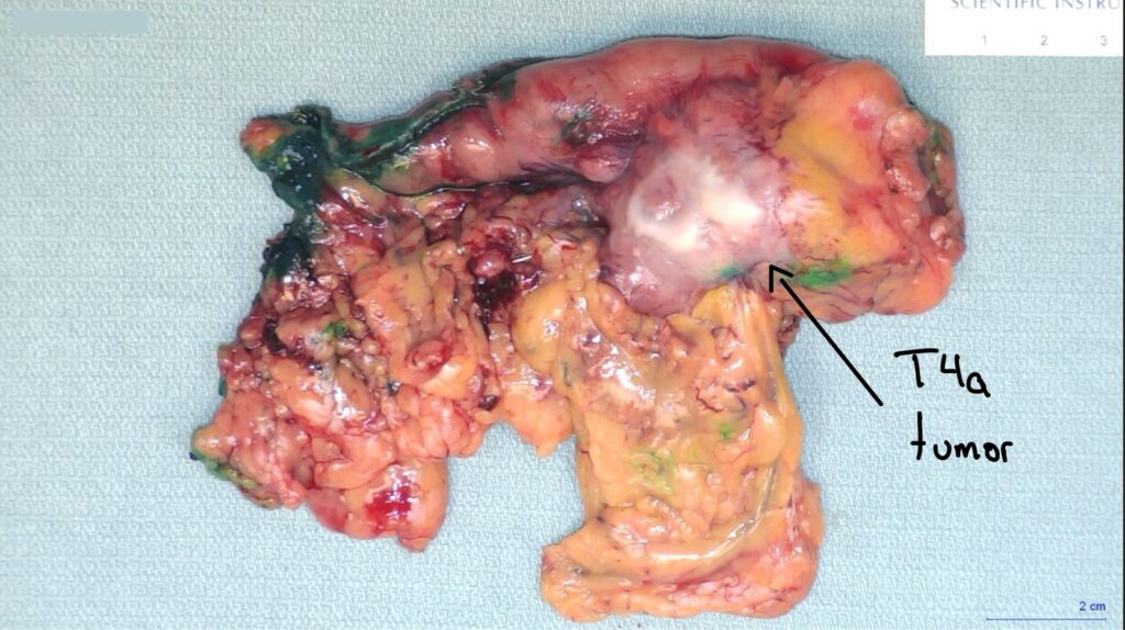
If the tumor has extended entirely through the muscularis/subserosa and extends to the serosal surface, it is a T4 tumor. T4a Includes tumor on the serosal surface, while T4b is reserved for tumors directly extending into an adjacent structure.
Take note of any serosal nodules or other adherent structures on the serosal surface of the stomach if present.
https://documents.cap.org/documents/Stomach_4.4.0.0.REL_CAPCP.pdf?_gl=1*9cq86q*_ga*MTc2OTc4NjM4MS4xNzQ3MzEzNjQw*_ga_97ZFJSQQ0X*czE3NTMyODQ0NjQkbzQkZzAkdDE3NTMyODQ0NjkkajU1JGwwJGgw
- Secondary pathology/background mucosa
After assessing the primary pathology, depth of invasion and distance to the margins, turn your attention to the background mucosa to look for any secondary pathologies (e.g. do the gastric folds appear thickened or effaced?)
Sample grossly normal areas, targeting the different regions of the stomach (cardia, fundus, body, antrum) to fully represent all the different histologic tissue types.
- Lymph node dissection
Once complete, proceed to a lymph node dissection. You need 16 lymph nodes for a gastrectomy specimen.
According to the CAP staging protocols, all of the nodes found along the lesser curve, the greater curve, the supra-GEJ nodes, and the infra-pyloric are all considered regional nodes. This is essentially all the fat that is received on a total gastrectomy specimen. Therefore there is no need to separate the nodes found from the lesser and greater omentum.
Example Gross Description
Patient and specimen identifiers. Received fresh/in formalin.
The specimen consists of a total gastrectomy which includes distal esophagus (1.3 cm in length with an open circumference of 3.0 cm at the proximal margin), stomach (10.5 cm in length along the lesser curve, 18.5 cm in length along the greater curve with an open circumference of 4.0 cm at the distal margin), lesser omentum (8.2 x 4.2 x 1.5 cm) and greater omentum (42.5 x 18.5 x 2.5 cm). A staple line is identified along the lesser omental margin (3.5 cm in length).
A smooth, firm, light tan area of puckering is identified on the anterior aspect of the gastric serosa. Opening reveals a raised, exophytic tan/necrotic tumor (10.2 cm proximal to distal x 5.2 transversely x 2.2 cm in thickness) located within the cardia and fundus of the stomach which extends along the anterior wall of the greater curve which is grossly consistent with the previously described area of puckering. Sectioning reveals tumor to invade through the muscularis, extending to the serosal surface at the previously described area of puckering. Tumor comes to the 5.2 cm of the proximal margin, 13.4 cm of the distal margin, 5.2 cm of the lesser omental margin, 6.0 cm of the staple line on the lesser omentum and 22.5 cm of the greater omental margin. No tumor identified within the lesser or greater omental fat.
The remaining uninvolved mucosa is grossly unremarkable. 21 possible lymph nodes identified with range in size from 0.3 to 0.8 cm greatest dimension. Photos are taken.
Inking:
Blue – lesser and greater omental margins
Green – serosa overlying tumor
Black – distal
Representative sections as follows:
A1-A2. Proximal margin, shave
A3-A6. Distal margin, shave
A7-A12. Contiguous section of tumor submitted from proximal to distal
A13-A14. Tumor extending to serosal surface
A15-A16. Lesser omental margin closest to tumor
A17-A18. Greater omental margin closest to tumor
A19. Vessels underlying staple line on lesser omentum, en face
A20. Gastroesophageal junction
A21-A24. Grossly normal mucosa (cardia, fundus, body, antrum respectively)
A25-A45. One possible lymph node bisected in each cassette
Pingback: Pathologists’ Assistants: What They Do - Canadian Path Assistant