Introduction: Why Depth of Invasion Matters
When doctors stage cancer, they assign a T stage based on how large the tumor is or how deeply it has grown into surrounding tissue.
In colon cancer, T staging is based on depth of invasion, not size.
The deeper a tumor grows into the bowel wall, the higher the T stage. A higher T stage usually means the tumor is more advanced and may require more aggressive treatment.
In simple terms:
- Size tells us how big the tumor is
- Depth tells us how far the tumor has grown into or through the bowel wall
For colon cancer, depth is what matters most.
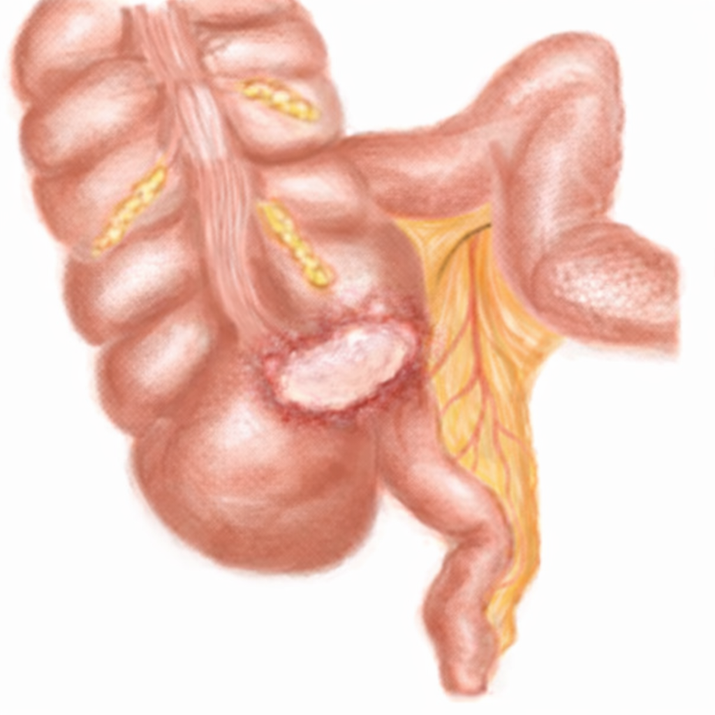
Quick Review: Layers of the Colon Wall
To understand T staging, it helps to remember that the colon is made up of several layers. Think of it like a tube with layers from inside to outside.
From innermost to outermost:
- Mucosa
- Submucosa
- Muscularis propria (contains circular/circumferential and longitudinal muscle fibers)
- Subserosa / pericolic fat
- Serosa (where present)
As the tumor grows outward through these layers, the T stage increases.
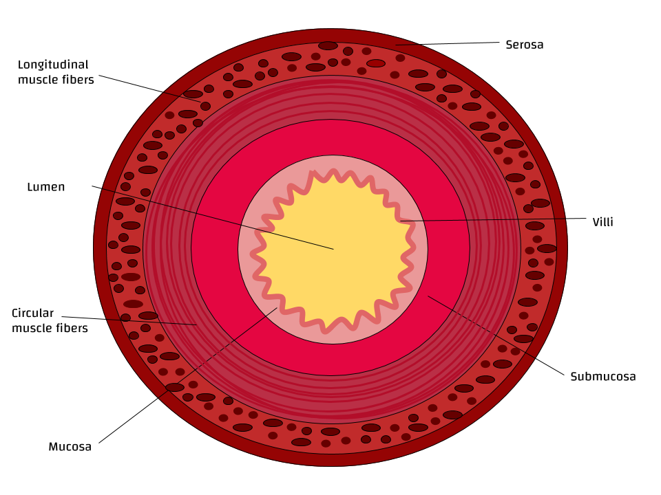
Jazlyn G, CC BY-SA 4.0 https://creativecommons.org/licenses/by-sa/4.0, via Wikimedia Commons
T1 Tumors: Invasion into Submucosa
A T1 tumor has grown through the mucosa and into the submucosa, but it has not reached the muscularis propria.
Grossly, this can be subtle. You may see a mucosal mass. When you section through it, the key question is:
Is the muscularis layer still intact and uninvolved underneath the tumor?
If there is a continuous, uninvolved muscularis layer beneath the lesion, the tumor is likely T1.
Gross Features
- Muscularis appears intact and continues beneath the tumor
- No obvious disruption of the muscle layer
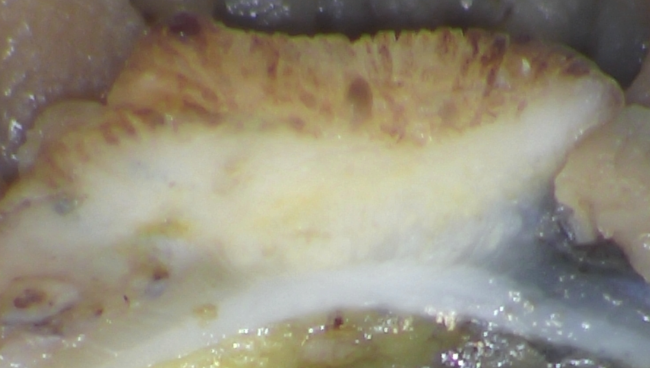
Key Takeaway
- T1 = tumor invades submucosa but not muscularis propria
T2 Tumors: Invasion into Muscularis Propria
A T2 tumor has grown from the mucosa into the muscularis propria, but it has not gone all the way through it.
At this stage, the tumor is involving the muscle layer.
Gross Features
- Tumor extends into the muscular layer
- The interface between tumor and muscle may look blurred
- However, a continuous muscularis layer still exists beneath the tumor
- No tumor is seen extending into pericolic fat
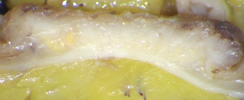
You should still be able to identify a clear boundary between tumor and fat.
Key Takeaway
- T2 = tumor invades into muscularis propria but does not extend beyond it
T3 Tumors: Invasion Through Muscularis into Pericolic Fat
A T3 tumor has grown completely through the muscularis and into the pericolic fat.
This is a major step up in staging.
Previously, the muscularis and fat were separated by a clear line. Once tumor invades through the muscle, that clean separation disappears.
Gross Features
- Loss of a distinct muscularis layer beneath the tumor
- Irregular white or tan projections extending into pericolic fat
- Blurred or disrupted muscle to fat interface
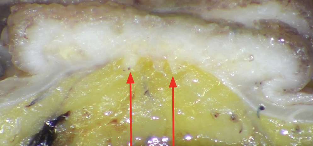
At this point, you can no longer identify an intact muscle layer separating tumor from fat. The red arrows show tumor extending into the fat.
Key Takeaway
- T3 = tumor extends through muscularis into pericolic tissue
T4 Tumors: Involvement of Serosa or Adjacent Structures
T4 tumors represent the most advanced local stage.
T4a: Tumor Reaches or Involves the Serosa
These tumors extend all the way through the bowel wall and reach the serosal surface.
Grossly, you may see:
- Tumor on the outer surface of the bowel
- Puckering of the serosa
- Serosal nodules
- Adherent tissue suggesting surface involvement
Important note: The rectum does not have a serosal covering along its distal portion. In those cases, T4a does not apply in the same way because that outer surface is a circumferential margin, not serosa.
T4b: Tumor Invades Adjacent Organs
This is when the tumor directly extends into nearby structures, such as:
- Small bowel
- Abdominal wall
- Other organs
Key Takeaway
- T4a = serosal involvement
- T4b = direct invasion into adjacent structures
Common Grossing Pitfalls That Affect T Staging
Small errors at the bench can lead to incorrect staging.
Common mistakes include:
- Not sectioning deeply enough through the tumor
- Not examining the entire tumor
- Assuming a tumor is T1 or T2 after only one section
Always section completely through the tumor until you see normal tissue beneath it. If you are unsure whether the tumor is confined to muscle or has entered fat, take additional sections.
When in doubt, submit more tissue.
Final Takeaway
Depth of invasion is one of the most important factors in colon cancer staging.
As a PA, your careful sectioning and sampling determine whether a tumor is staged as T1, T2, T3, or T4.
The key question to ask yourself every time is:
What is the deepest layer this tumor has reached?
If you answer that correctly and sample it well, you have done your job.